GOOD TRIPS 体彩官网实时结果®
A 168网结果查询记录+飞艇综合福彩开采查询网站 168官网极速体彩预测 Travel Studio for Hotel Lovers
Travel Made 幸运168中国-幸运飞行艇168体彩历史记录
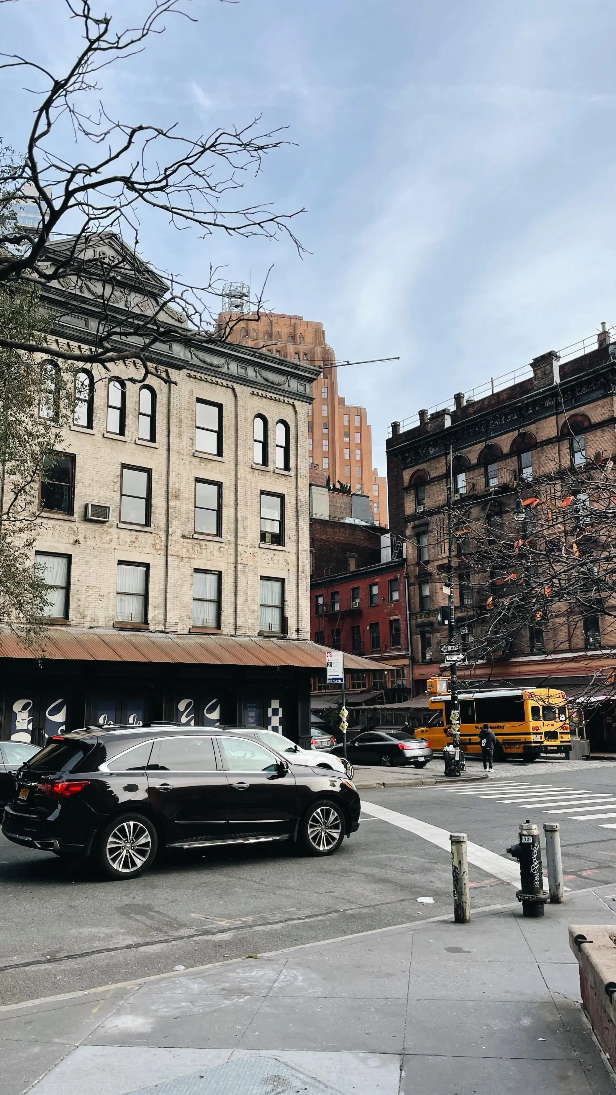
At Good Trips, travel is 168网最新开奖直播全国飞行艇统一开奖记录 with the same care as a well-built space: intentional, functional, and beautiful. Every trip starts with the right hotel — a place with soul, design integrity, and people who care—and expands outward to create an experience that feels natural and deeply personal.
Our clients don’t have time to guess which places are worth it. They trust us to curate travel that’s aesthetically inspiring, emotionally grounding, and 幸运的飞行艇168体彩网最新开奖记录、飞艇168号码结果查询 effortless. Because an investment in travel should always feel like an investment in yourself.

ABOUT GOOD TRIPS
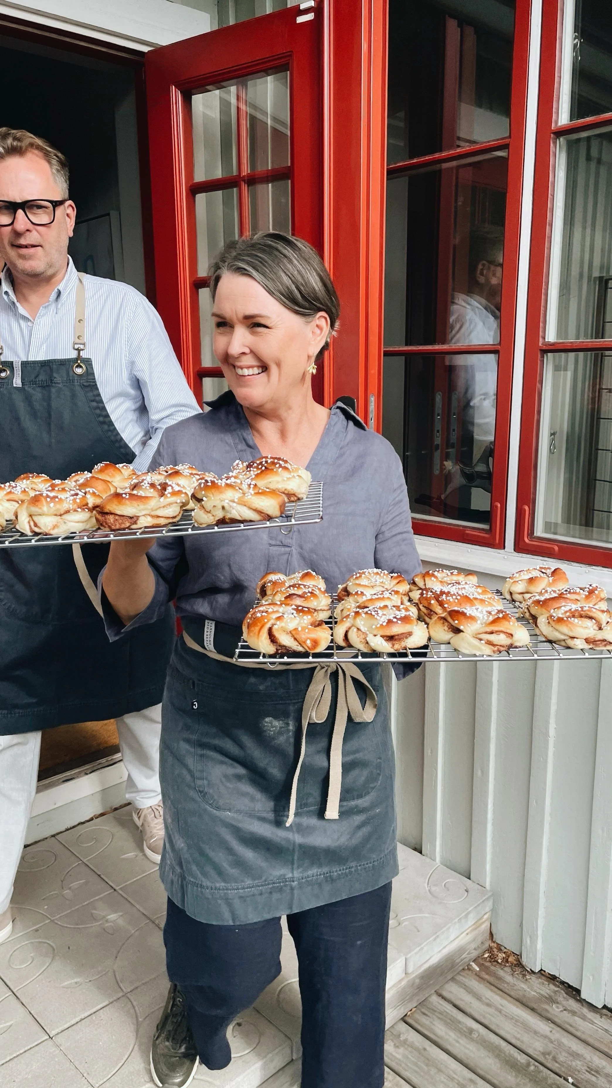
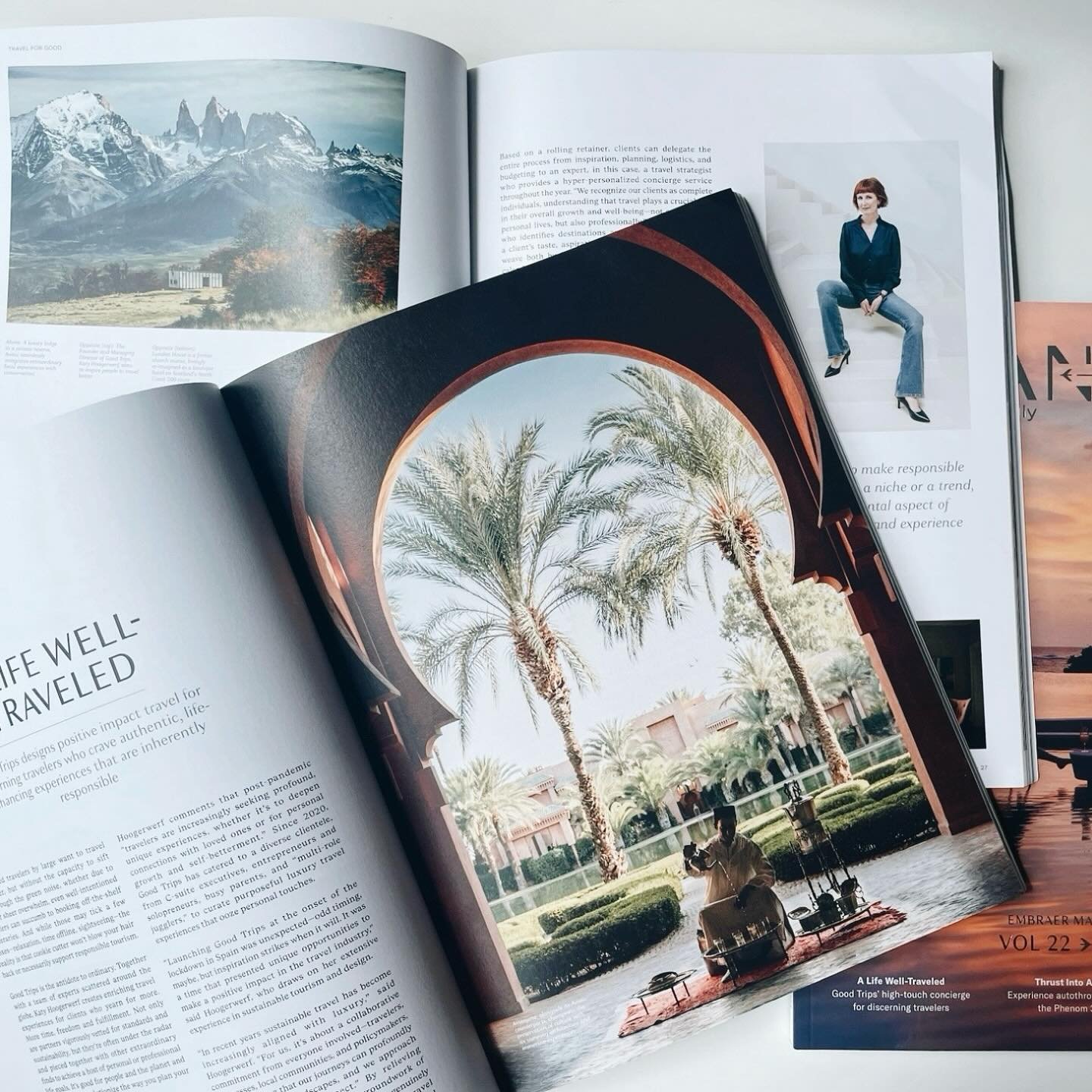
Good Trips began with a simple belief: the right hotel can define an entire trip. Founded by Katy Hoogerwerf, the studio merges design sensibility, wellness insight, and years of hospitality experience to create journeys that feel grounded, beautiful, and human.

OUR SERVICES
Whether you want a single stay handled beautifully or a full itinerary shaped around your life, Good Trips offers a considered approach to travel design. Each service — from Tailored Bookings to fully Curated Trips — is rooted in care, intuition, and an unshakable sense of style.
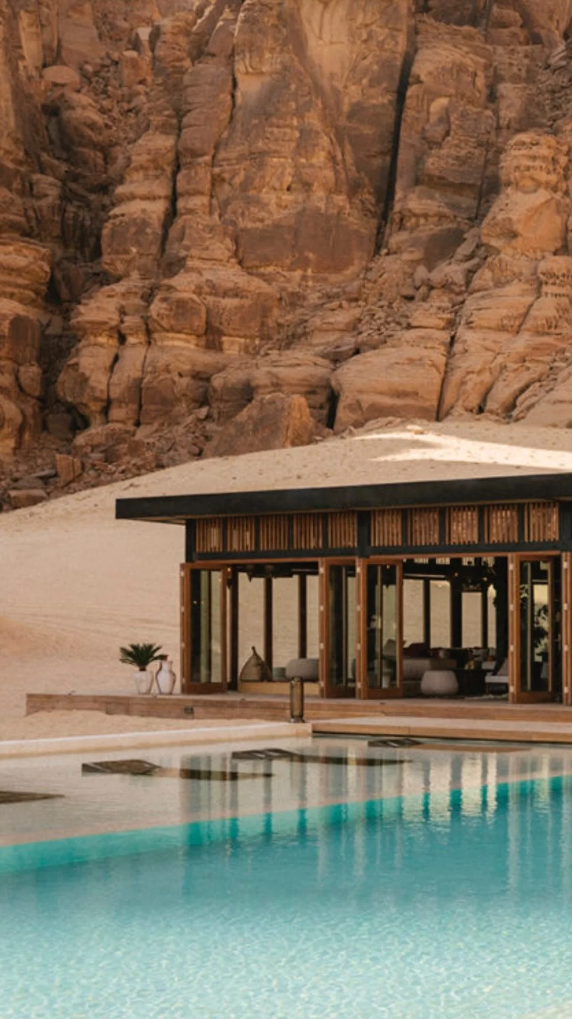
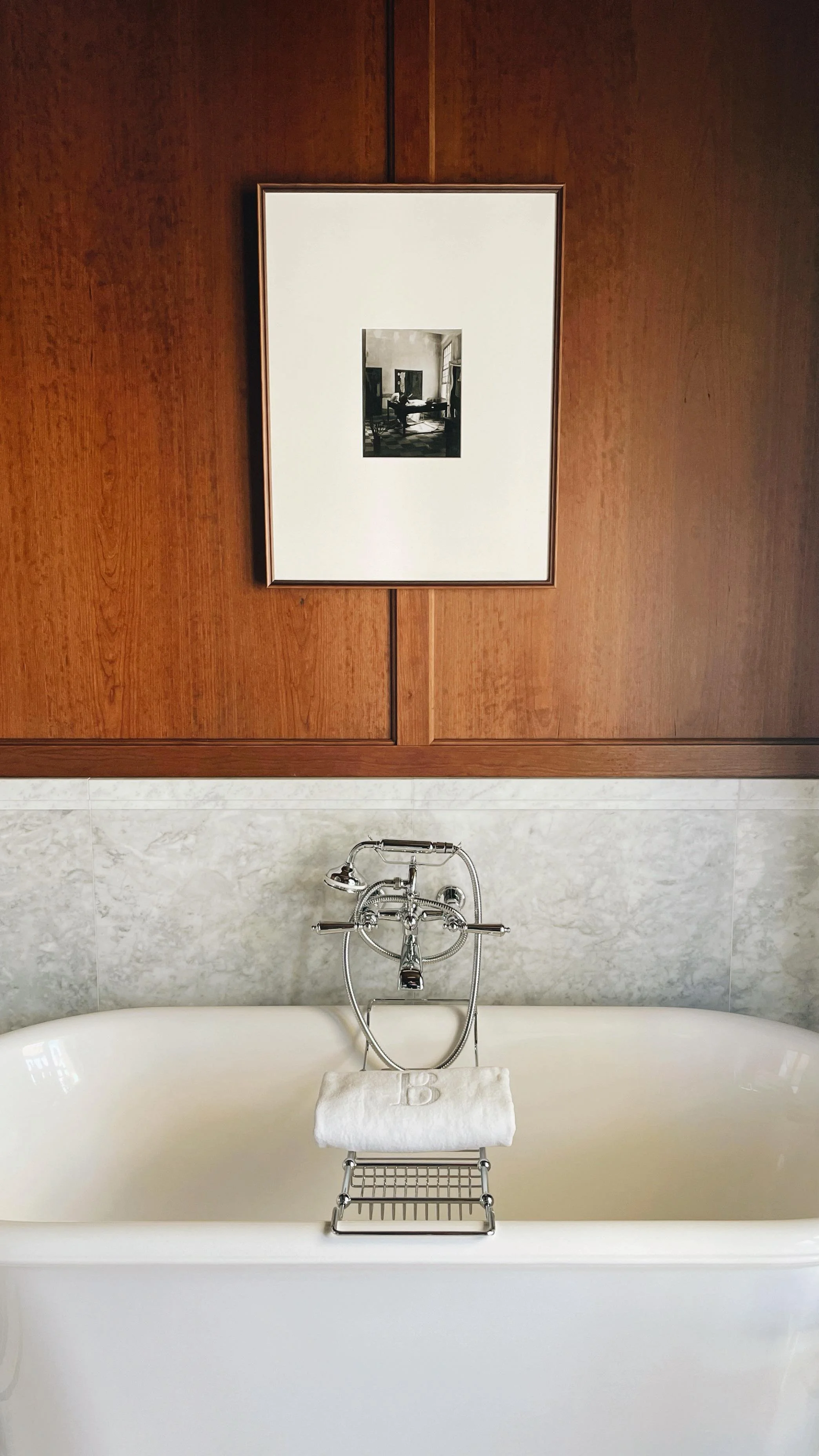
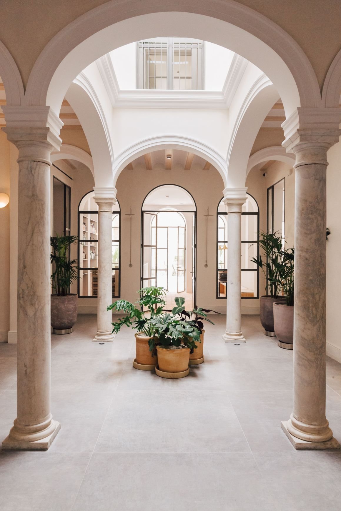
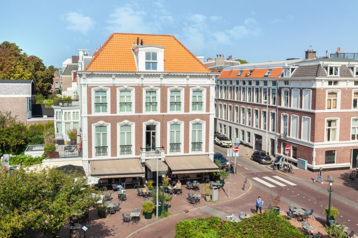
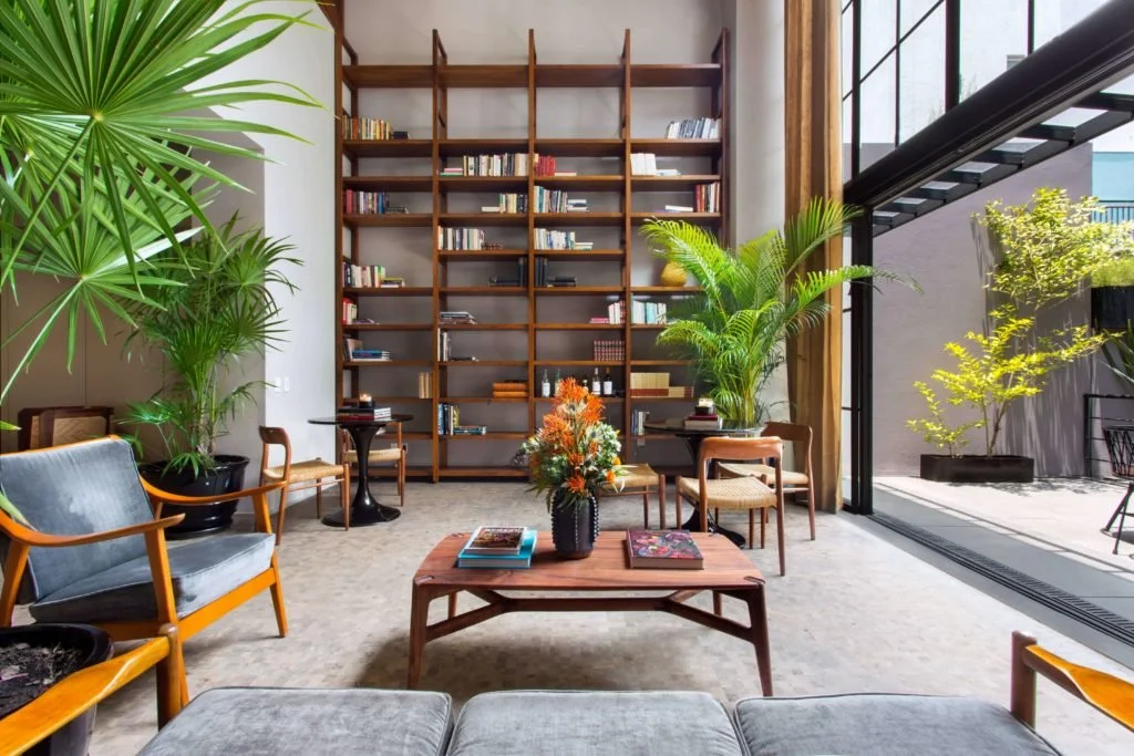
FEATURED HOTELS


Good Brands 幸运飞艇开采直播-中国飞行艇168幸运体彩预测网最新结果
The Good Brands Directory is 稳定168网免费查询和正规福彩平台 幸运168官方网站-体彩飞行艇实时结果 of our favorite travel essentials. Curated for design, durability, and 中国飞行艇168幸运体彩预测网最新, they’ll help you travel your best.

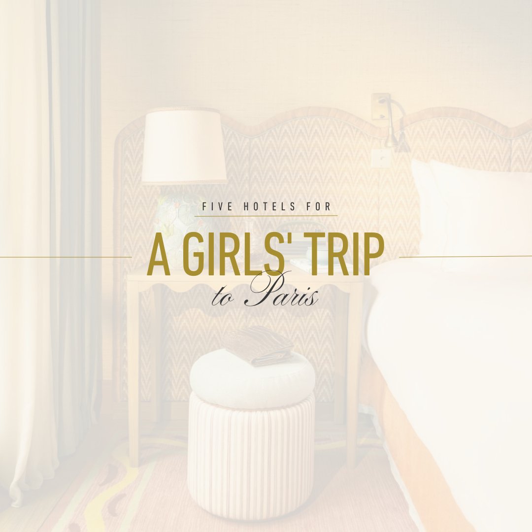
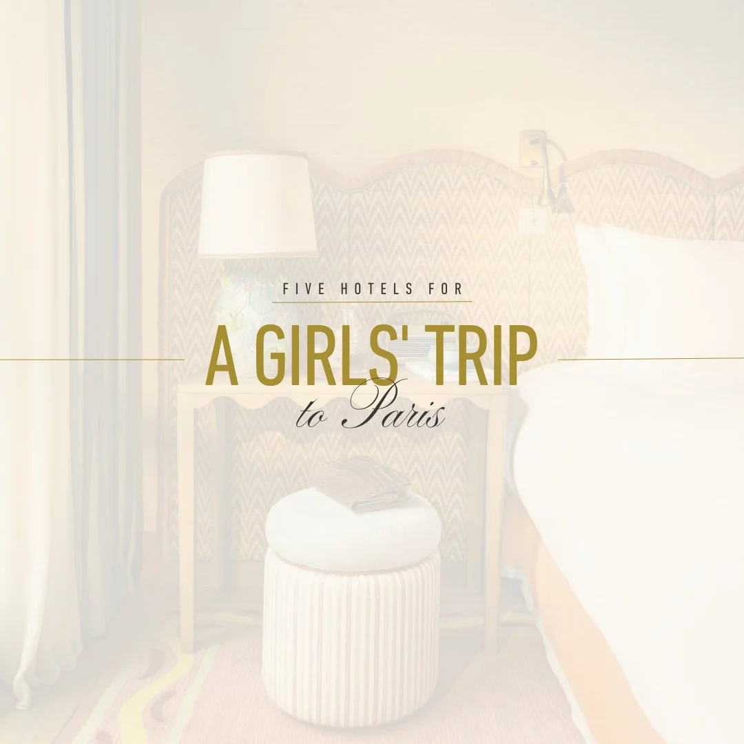
TRAVEL JOURNAL


Get Social